
The short answer
A diagram can show where neurons connect, and a fluorescent marker can show where a gene is expressed. Neither observation alone tells us which cells are required for a response. Chalfie’s work on touch in a transparent roundworm offers a way to compare these different questions.
Original source comparison
Match the claim to the observation
These are complementary records, not interchangeable proofs.
| Country | What the evidence supports | What it does not establish |
|---|---|---|
| Anatomy | A route exists in the reconstructed network | It carries every response in every developmental state |
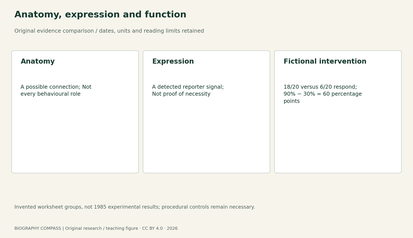
| Reporter signal | A construct produces a detected signal under stated conditions | The labelled cell is necessary or sufficient for behaviour |
| Intervention | A measured response differs after a specified change | All procedural and off-target alternatives have been excluded |
Chronology
A timeline of the turning points
Circuit paper
Anatomical and functional evidence were combined.
GFP idea
The later lecture dates the seminar that prompted a marker idea.
Retrospective lecture
Chalfie explains the research route and collaborators.
The historical question concerned a defined response
The 1985 paper The neural circuit for touch sensitivity in Caenorhabditis elegans names Chalfie, Sulston, White, Southgate, Thomson and Brenner. Its abstract describes six touch receptor neurons, five pairs of interneurons and 69 motor neurons involved in the response. These numbers concern the reported touch circuitry, rather than a count of every neuron in the animal. The study combined serial-section electron-microscope anatomy with selective removal of identified cells through precursor-cell intervention. Its importance here is the combination of structural and functional evidence. This guide consulted the original abstract, not the full experimental methods or every reconstruction.
Sources for this section: [1]
An anatomical connection is a route, not a complete explanation
A connection map identifies possible paths through which activity might travel. It does not, by itself, establish which route carries the response in a particular condition. The paper’s abstract describes changes during development, including additional electrical connections and use of a further interneuron pathway. A diagram therefore needs a developmental context as well as cell names. Treating one image as a timeless explanation of all behaviour would discard that qualification. The abstract supports a comparison of evidence types, but cannot supply all the controls or quantitative effects that a complete evaluation of the experiments would require.
Sources for this section: [1]
Expression answers a different biological question
Chalfie’s later Nobel lecture explains his work on mechanosensation and why older methods of marking gene expression were limiting. He recalls hearing about GFP in 1989 and contacting Douglas Prasher about the gene. A fluorescent expression marker could help identify where a genetic construct was active in a living transparent animal. That observation is useful without being identical to a functional intervention. A signal in a neuron does not alone establish that the neuron is necessary for a behaviour. Likewise, lack of visible signal can reflect the construct, detection conditions or timing, rather than conclusively establishing that a biological process is absent.
Sources for this section: [2]
Original exercise: assign each claim to its evidence
Consider three fictional records about a cell called N. Record A is an anatomical drawing showing a connection from N to a motor pathway. Record B shows a reporter signal in N under the stated imaging conditions. Record C compares 20 untreated animals, of which 18 respond to touch, with 20 animals after an intervention targeting N, of which 6 respond. These are invented teaching observations, not the 1985 results. Calculate the observed response proportions and their difference. Match each record to a structural, expression or functional claim. Then identify a control needed before treating the difference as an effect specifically attributable to N.
Reference answer: an intervention still needs a comparison
The observed proportions are 18/20 = 90% and 6/20 = 30%, a difference of 60 percentage points. Record A supports the stated anatomical connection; B supports detection of the reporter under those conditions; C associates the intervention with a lower observed response. A sham-treated comparison would help distinguish targeting N from effects of handling or the procedure. Checks on off-target injury, assignment, scoring and the animal’s general capacity to move would also matter. The two small groups alone do not establish a universal effect size, the complete sensory circuit or sufficiency: showing that activity in N alone can trigger the response is another experimental question.
Sources for this section: [1]
Read the retrospective beside the original abstract
The lecture explains a researcher’s later account of motivation and collaborators, while the abstract identifies what a specific paper reports. Put each claim beside the record that can support it. The lecture’s account of a 1989 idea should not be used as the publication date of the 1985 circuit study, and the later usefulness of GFP should not be projected backwards as the method used for every earlier experiment. The practical reading habit is to separate what was connected, what was observed, what was changed and which response was measured. The linked GFP guide follows a different question: how a protein became a genetic marker.
Continue the chronology
Related history guides
Tsien’s Calcium Indicators: Why a Ratio Needs Calibration
Compare a different measurement question and its explicitly bounded worked exercise.
Read the timeline1985–1992 research; 2014 participant lecturesBlue LEDs: GaN Growth and p-Type Conduction Were Different Barriers
Compare a different measurement question and its explicitly bounded worked exercise.
Read the timeline1961–1994 researchGFP: From an Isolated Protein to a Fluorescent Marker
Follow the separate question of isolation, cloning and expression.
Read the timelineBiography context
Explore United States through connected lives
Connected biographies
People behind the work

Born 1947
Martin Chalfie
Molecular biologist and neurobiologistMartin Chalfie is an American biologist whose studies of a transparent roundworm helped him recognise the value of green fluorescent protein as a marker. His laboratory’s experiments connected a jellyfish protein with the ability to investigate gene expression in living organisms.Read biographyBorn 1951
Douglas Prasher
Molecular biologistDouglas Prasher is an American molecular biologist whose cloning and sequence work supplied an essential step in the development of green fluorescent protein as a research tool. His papers and the accounts of later collaborators make his contribution visible beyond the list of Nobel laureates.Read biographyQuick answers
Common questions
- Are the 20-animal groups historical data?
- No. They are invented worksheet observations, clearly separated from the paper abstract.
- Does this guide reproduce a cell-ablation protocol?
- No. It teaches how to interpret evidence and controls, not how to perform an intervention.
Primary and institutional sources
How this history was checked
Compare the identified original texts, participant accounts and institutional records; the teaching examples are separately labelled. This article was reviewed on 27 October 2026.
- Martin Chalfie, John Sulston, John White, Eileen Southgate, J. Nicol Thomson and Sydney Brenner; Europe PMCThe neural circuit for touch sensitivity in Caenorhabditis elegans,JNeurosci5(4),956–964,1985: original abstract
- Martin Chalfie; Nobel FoundationGFP: Lighting Up Life,8 December 2008,150–169
- Martin Chalfie; Nobel FoundationMartin Chalfie: autobiographical account,2008