
The short answer
How did a protein isolated from jellyfish become a tool for studying cells? The answer involves separate chemical and genetic achievements. This guide compares the records for those steps, then uses clearly invented readings to show why a glowing sample still needs controls.
Original source comparison
What kind of claim is being made?
Assign each claim to the record that can support it.
| Country | What the evidence supports | What it does not establish |
|---|---|---|
| Participant lecture | A retrospective chronology and account of chemical work | Full independent replication of every cited paper |
| 1994 abstract | Reported expression and fluorescence in named systems | Every later organism, construct or target localisation |
| Teaching readings | 100 units after stipulated background subtraction | Historical measurements or proof of protein function |
Chronology
A timeline of the turning points
Isolation and publication
The participant account separates early work from its publication.
Chromophore investigation
Chemical work addressed the fluorescent structure.
Cloning
Prasher and colleagues are credited in the lecture.
Expression
The original abstract identifies bacteria and nematode cells.
Isolation and genetic expression answer different questions
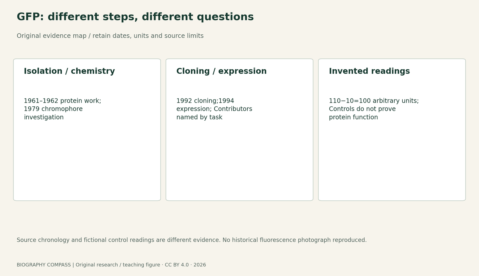
Osamu Shimomura’s 2008 lecture describes GFP as a byproduct of work on aequorin in 1961, with the early research published in 1962. Isolating a fluorescent substance establishes that it can be obtained and investigated; it does not show that another organism can make it from introduced DNA. The later ability to express GFP therefore needed additional work. Treat discovery, publication and application as separate entries in a research ledger. The biographical account and MBL obituary also name collaborators and Akemi Shimomura’s practical contributions, which a single-inventor timeline can easily lose.
Aequorin’s light and GFP fluorescence are not identical
In the jellyfish system, the lecture describes energy from aequorin-associated blue light being transferred to GFP, which emits green light. GFP is fluorescent: an excitation source supplies energy before the emission is observed. Calling it a marker that glows can be convenient shorthand, but it should not imply that a purified sample supplies unlimited light without excitation. The distinction matters when reading a photograph or designing a control. The appearance of a green signal establishes an observation under particular conditions; identifying the molecular system responsible requires further evidence.
Sources for this section: [1]
The 1979 chemistry preceded the 1990 s genetic work
Shimomura’s lecture explains his investigation of the chromophore within GFP’s peptide chain and dates that work to 1979. It then credits Prasher and colleagues with cloning GFP in 1992 and Chalfie and colleagues with expression in living organisms in 1994, followed by further development associated with Tsien. The consulted lecture is a participant’s later synthesis, not the complete methods of every earlier paper. Its chronology is useful precisely because it distinguishes tasks. Finding a protein, studying its chemical structure, obtaining the coding sequence and demonstrating a biological application are connected contributions rather than interchangeable descriptions of one experiment.
Sources for this section: [1]
The original 1992 abstract supplies a separate record
Prasher, Eckenrode, Ward, Prendergast and Cormier’s 1992 Gene paper reports cloning and sequencing GFP complementary DNA and genomic material. Its abstract identifies a cDNA encoding a 238-amino-acid polypeptide. That is a concrete molecular result, not the same claim as demonstrating fluorescence after expression in bacteria and worms in the later Science report. Prasher appears on both author lists. The two abstracts therefore identify connected but distinct contributions. Only the original abstract and bibliographic fields of the 1992 paper were consulted here; its complete methods, figures and all expression tests were not acquired.
Sources for this section: [5]
What the original 1994 abstract actually reports
The Science abstract names Chalfie, Tu, Euskirchen, Ward and Prasher. It reports a fluorescent product after GFP complementary DNA was expressed in Escherichia coli and Caenorhabditis elegans cells. It also states that added substrates and cofactors were not required for that fluorescence. This supports use as a marker of expression and localisation, but the abstract alone is not a full account of imaging conditions, every construct or every possible organism. It does not make an ordinary brightness reading a universal measure of protein function. This guide consulted the original abstract and bibliographic fields through Europe PMC, rather than claiming to inspect all of the paper’s experimental figures.
Sources for this section: [3]
Original worksheet: separate signal from background
Use these invented fluorescence readings in arbitrary units: A, no GFP and excitation on, 10; B, GFP and excitation on, 110; C, GFP and excitation off, 12. Assume the same detector settings and measurement interval. Calculate B minus A. Explain why B compared only with C tests the effect of excitation in the stated GFP-containing sample, rather than the effect of adding GFP: both B and C contain GFP, and only excitation differs. Then decide whether the corrected signal establishes where a particular target protein is located, if no target-specific fusion or localisation control is described. Finally place 1961 isolation work, 1962 publication, 1979 chemistry, 1992 cloning and 1994 expression in a table whose last column names the scientific question answered. The readings are editorial teaching data, not recovered results from the historical papers.
Reference answers: what the controls allow
The background-corrected reading is 110−10=100 arbitrary units. A and B share the excitation condition and differ in the stated GFP condition. B and C both contain GFP and differ only in excitation, so their comparison addresses excitation rather than GFP presence. The information supplied does not establish a target protein’s location or biological function. A stronger claim would require the relevant construct, controls and imaging evidence. In the chronology, isolation asks what material can be separated, chromophore chemistry asks what produces fluorescence, cloning concerns the coding sequence, and expression tests whether another cellular setting can produce a fluorescent product. The 1962 entry dates publication rather than a fifth scientific task. This is the central historical lesson: a new use became possible through distinct contributions, each with its own evidential job.
Continue the chronology
Related history guides
Esaki’s Tunnel Diode: Reading Negative Differential Resistance
Compare another discovery through its measured quantity and a separately labelled teaching model.
Read the timeline1985 circuit paper; 2008 participant lectureChalfie’s Worm Research: Anatomy, Expression and Function
Compare a distinct biological measurement question through its cited records and original worksheet.
Read the timeline1985 calcium-indicator paperTsien’s Calcium Indicators: Why a Ratio Needs Calibration
Compare a distinct biological measurement question through its cited records and original worksheet.
Read the timelineBiography context
Explore United States through connected lives
Connected biographies
People behind the work

1928–2018
Osamu Shimomura
Biochemist and bioluminescence researcherOsamu Shimomura was a Japanese chemist who isolated proteins involved in jellyfish light production. His work on aequorin and green fluorescent protein, GFP, helped provide the chemical foundation for tools that later researchers developed to study living cells.Read biographyBorn 1951
Douglas Prasher
Molecular biologistDouglas Prasher is an American molecular biologist whose cloning and sequence work supplied an essential step in the development of green fluorescent protein as a research tool. His papers and the accounts of later collaborators make his contribution visible beyond the list of Nobel laureates.Read biography
Born 1947
Martin Chalfie
Molecular biologist and neurobiologistMartin Chalfie is an American biologist whose studies of a transparent roundworm helped him recognise the value of green fluorescent protein as a marker. His laboratory’s experiments connected a jellyfish protein with the ability to investigate gene expression in living organisms.Read biography
1952–2016
Roger Y. Tsien
Chemist and biological-imaging researcherRoger Y. Tsien was an American chemist who designed molecules to investigate activity inside cells. His research ranged from calcium indicators to improved fluorescent proteins, connecting chemical structure with the kinds of biological questions that researchers could measure.Read biographyQuick answers
Common questions
- Does the guide reproduce the1994 microscopy experiment?
- No. It compares an original abstract with a later lecture and supplies separately labelled invented readings.
- Does fluorescent expression necessarily prove genomic integration?
- The consulted abstract reports expression from complementary DNA. This guide does not infer a particular integration event or construct beyond that statement.
Primary and institutional sources
How this history was checked
Compare the identified original texts, participant accounts and institutional records; the teaching examples are separately labelled. This article was reviewed on 27 October 2026.
- Osamu Shimomura; Nobel FoundationDiscovery of Green Fluorescent Protein, GFP: Nobel Lecture,8 December 2008,117–129
- Marine Biological LaboratoryOsamu Shimomura: institutional obituary,19 October 2018
- Martin Chalfie, Yuan Tu, Ghia Euskirchen, William W. Ward and Douglas C. Prasher; Europe PMC bibliographic record,PMID8303295Green fluorescent protein as a marker for gene expression, Science 263(5148),802–805,1994: original abstract
- Nobel FoundationOsamu Shimomura: prize facts
- Douglas C. Prasher, Virginia K. Eckenrode, William W. Ward, Franklyn G. Prendergast and Milton J. Cormier; Europe PMCPrimary structure of the Aequorea victoria green-fluorescent protein, Gene111(2), 229–233,1992: original abstract