
The short answer
How did pulse–chase autoradiography help connect intracellular compartments without directly filming every protein molecule?
Read the evidence
Different records within the same investigation
The following comparison distinguishes the specific questions answered by the inspected records.
| Record or test | What it establishes | What remains different |
|---|---|---|
| Micrograph | Provides spatial organisation in a preparation. | Does not alone supply a time sequence. |
| Pulse–chase distribution | Tracks a labelled cohort across intervals. | Is not continuous identification of every molecule. |
| Fractionation | Provides material and quantitative distributions. | Requires careful assignment of compartments. |
The question required time information as well as structure
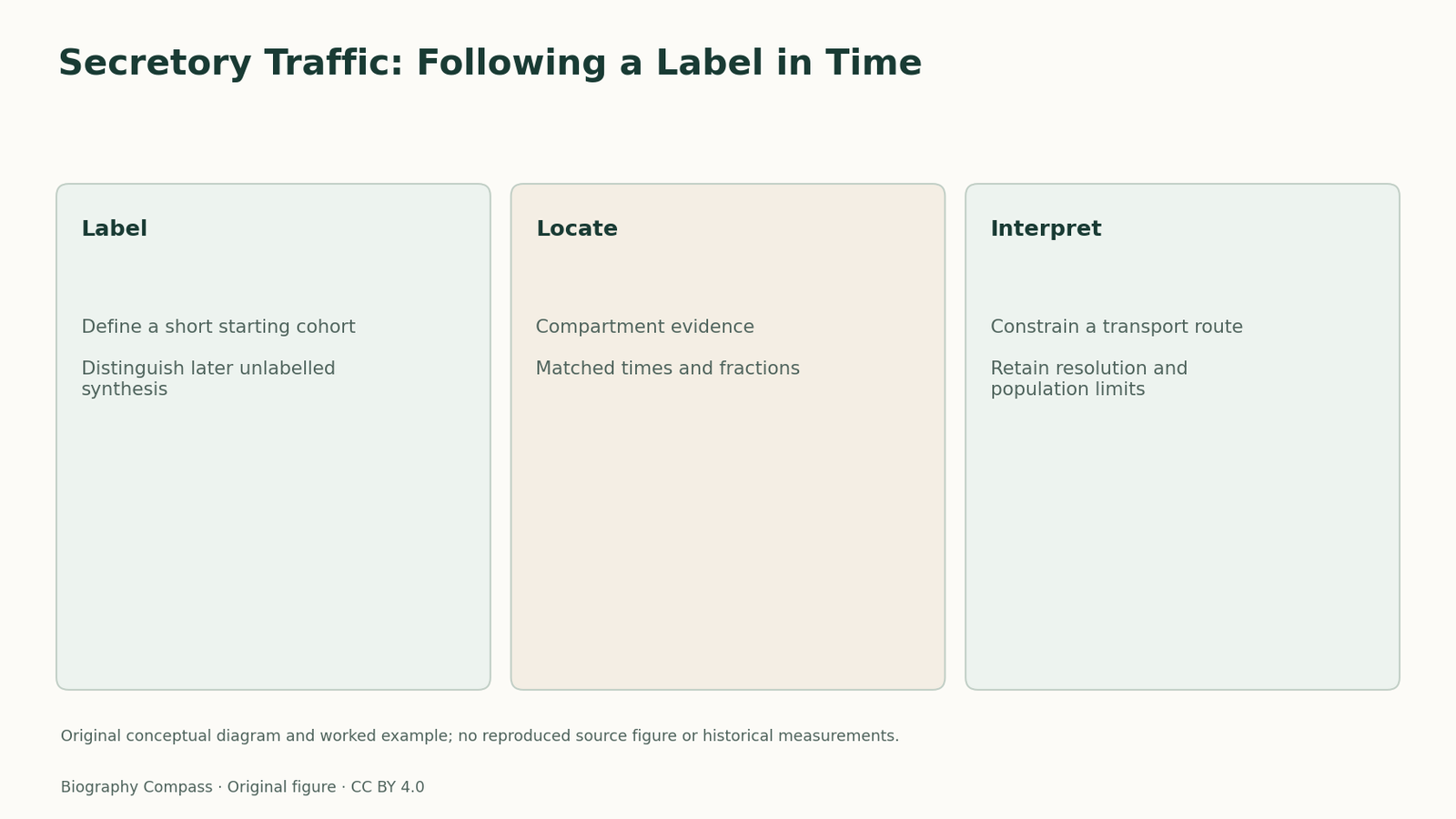
A micrograph can reveal cellular compartments without showing how a newly made protein moves among them. The Jamieson–Palade programme asked how secretory material travelled through pancreatic exocrine cells. Its 1967 studies combined labelled amino-acid incorporation with subsequent observation of intracellular locations. The experimental advantage was the ability to distinguish a relatively brief starting cohort from continuously added label. That made a temporal comparison possible without claiming that the investigators watched each protein individually cross every boundary.
A pulse and a chase separated stages of the inference
In Palade’s lecture, a brief exposure to label followed by removal of unincorporated label improved time resolution. The chase does not mean that all proteins disappear or that the cell stops making proteins. It changes the labelling history so that the distribution of an earlier cohort can be followed more meaningfully. This distinction matters when interpreting a later signal: it concerns where labelled material is found after an interval, not simply where all proteins in the cell happen to be most abundant.
Sources for this section: [2]
Fractionation and radioautography answered complementary questions
The original 1967 work investigated microsomal fractions and the Golgi periphery, while microscopy supplied spatial evidence. Palade’s autobiographical account distinguishes biochemical approaches from radioautographic studies with Caro and Jamieson. The methods constrain each other: a labelled fraction needs a defensible cellular assignment, and an image benefits from information about the labelled material and its timing. Neither should be treated as an automatically pure or perfectly resolved representation of an entire compartment. The pathway emerges from the combined pattern, rather than one isolated labelled spot.
A pathway model still had unresolved mechanisms
The Nobel lecture explicitly retained open questions about transport and the geometry of compartment connections. This is more informative than retroactively giving the historical experiments every detail of a later molecular model. The observed sequence supported movement through the secretory system in the investigated cells; it did not by itself identify every energy-requiring reaction or transport force. The wider Nobel award recognised cell organisation, but its broad wording is not a substitute for stating which preparation, labelled population and observation supported this particular pathway inference.
An original timing exercise and answer
Imagine an invented labelled cohort that is initially concentrated in compartment A, later in B and later still in C. Does the sequence alone prove that every molecule followed A–B–C with no alternatives? No. It is consistent with that route, but population measurements can conceal branching, incomplete recovery or different transit times. These letters and observations are fictional, not a historical table. A stronger reading asks whether the same labelled material is being compared, how compartments were identified and whether intervening pools could have been missed. The supported answer uses timing to constrain a route while keeping measurement resolution visible. It also distinguishes the experiment from a live film in which individual molecules are continuously identified across the full journey.
Continue the chronology
Related history guides
Cell Fractionation: A Pellet Is Not Purity
Examine Claude’s liver-cell fractionation studies through operational separation, biochemical activity and microscopy, with an original enrichment-versus-purity exercise.
Read the timeline1951–1974Lysosomes 1955: Why Enzyme Activity Was Hidden
Examine de Duve’s lysosome inference through latent activity, distribution patterns and later structural evidence, with an original accessibility-versus-absence exercise.
Read the timeline1971–1999Protein Targeting 1975: Timing the Membrane
Compare Blobel and Dobberstein’s 1975 transfer studies through precursor processing, protected location and different products, with a membrane-transfer exercise.
Read the timelineBiography context
Explore United States through connected lives
Connected biographies
Researchers and related approaches
1912–2008
George E. Palade
Cell biologist and microscopy researcherGeorge Palade combined microscopy, fractionation and labelling studies to investigate how secretory proteins move through cellular compartments.Read biography
1952–
Venkatraman Ramakrishnan
Structural biologistVenkatraman Ramakrishnan moved from physics to molecular biology and helped determine ribosomal structures, sharing the 2009 Nobel Prize in Chemistry.Read biographyQuick answers
Common questions
- Does a chase stop all protein synthesis?
- No. It changes the history of the label being followed.
- Was the experiment a live molecular film?
- No. Time-resolved population and spatial evidence supported the pathway interpretation.
Primary and institutional sources
How this history was checked
The references below distinguish the original study from later explanations. The worked exercise and comparison are newly written for this article. This article was reviewed on 8 November 2026.
- James D. Jamieson and George E. Palade, Journal of Cell Biology, 1967Intracellular Transport of Secretory Proteins in the Pancreatic Exocrine Cell. I. Role of the Peripheral Elements of the Golgi Complex
- Nobel Prize OutreachGeorge E. Palade — Nobel lecture
- Nobel Prize OutreachGeorge E. Palade — Facts
- Nobel Prize OutreachGeorge E. Palade — Biographical