
The short answer
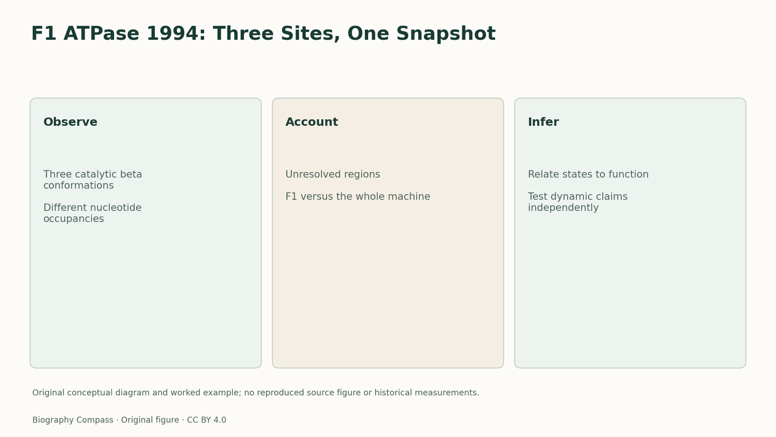
What did the three catalytic-site conformations reveal, and why was a static structure not itself a rotation movie?
Read the evidence
Different records within the same investigation
The following comparison distinguishes the specific questions answered by the inspected records.
| Record or test | What it establishes | What remains different |
|---|---|---|
| Different conformations | Describe structural asymmetry among catalytic subunits. | Do not provide a directly filmed reaction sequence. |
| Unresolved component | May be present but lack a reliable atomic model. | Should not automatically be called absent. |
| Rotation mechanism | Combines structural and functional constraints. | Cannot be established by the static snapshot alone. |
The experiment asked how catalytic sites were organised
The 1994 Nature report presented the structure of bovine mitochondrial F1-ATPase at 2.8-ångström resolution. F1 is a catalytic portion of the wider ATP-synthase machinery. The concrete question is what its subunits and bound nucleotides revealed about possible catalytic states. Naming the portion matters: a result about F1 should not be described as though it resolved the entire membrane-associated machine. Likewise, the reported resolution describes the structural measurement, not a temporal resolution for watching an enzyme operate.
Three beta subunits were not structurally equivalent
Abrahams, Leslie, Lutter and Walker reported differences among the three catalytic beta subunits in conformation and nucleotide occupancy. This asymmetry supplied a way to connect distinct structural states with a catalytic cycle. A simple picture of three permanently equivalent sites would miss the main observation. However, assigning a sequence of events to a set of different conformations remains an interpretation informed by biochemical evidence. The structure itself does not record a timestamped transition from one state to the next.
Sources for this section: [1]
The model also had explicit unresolved parts
The MRC structural history notes that delta, epsilon and parts of gamma were unresolved in the 1994 structure, although the subunits were present in the crystals. Failure to resolve a component therefore does not mean that it was biologically absent. Later structures added information. This provides a practical reading rule for structural claims: distinguish the material present in a sample from the part represented reliably in the final atomic model. A model can reveal a decisive organisational feature while remaining incomplete in other regions.
Sources for this section: [2]
Boyer and Walker supplied complementary constraints
Walker’s Nobel recognition was shared with Boyer for the mechanism of ATP synthesis. Boyer’s binding-change research addressed differences in chemical behaviour and energy coupling, while the F1 structure supplied a spatial arrangement consistent with distinct catalytic states. The connection does not turn a crystal into a direct rotation experiment. It shows how independent forms of evidence can narrow a mechanism together. Walker’s autobiography also places the programme within protein chemistry and mitochondrial sequence work, making the path from component identification to structural interpretation visible.
An original snapshot exercise and answer
Imagine a fictional machine photographed once with three similar stations: one empty, one holding an input and one holding a product. What can the image establish? It supports the observation that the stations differ in that captured arrangement. It does not, by itself, prove their time order, the direction of movement or the speed of the machine. These are teaching stations, not labels copied from the F1 paper. A student could propose a cycle consistent with them, but would need independent evidence to distinguish that cycle from alternatives. The answer should also ask whether a missing part was absent from the sample or merely unresolved in the image. This separates observed asymmetry, dynamic interpretation and model completeness.
Continue the chronology
Related history guides
ATP Synthase: Binding, Chemistry and Release
Read Boyer’s 1973 working hypothesis beside the 1976 binding study, distinguishing ATP formation, substrate commitment and energy-dependent product release.
Read the timeline1961–1978Chemiosmosis: More Than a Proton Count
Examine Mitchell’s chemiosmotic framework through chemical and electrical contributions, with a worked exercise separating a pH difference from total driving force.
Read the timelineBiography context
Explore United Kingdom through connected lives
Connected biographies
Researchers and related approaches
1941–
John E. Walker
Biochemist and structural researcherJohn Walker connected protein chemistry with ATP-synthase structure, leading research that revealed different catalytic conformations in bovine mitochondrial F1.Read biography
1918–2013
Frederick Sanger
Biochemist and sequencing-method researcherFrederick Sanger developed ways to determine the order of components in proteins and nucleic acids. His two chemistry Nobel Prizes mark different stages of a research career whose continuity was the problem of sequence and whose methods depended on many collaborators.Read biographyQuick answers
Common questions
- Was the 1994 structure the entire ATP synthase?
- No. It concerned bovine mitochondrial F1 and included unresolved regions.
- Did crystallography directly film rotation?
- No. It supplied a static structural model that supported mechanistic interpretation.
Primary and institutional sources
How this history was checked
The references below distinguish the original study from later explanations. The worked exercise and comparison are newly written for this article. This article was reviewed on 8 November 2026.