
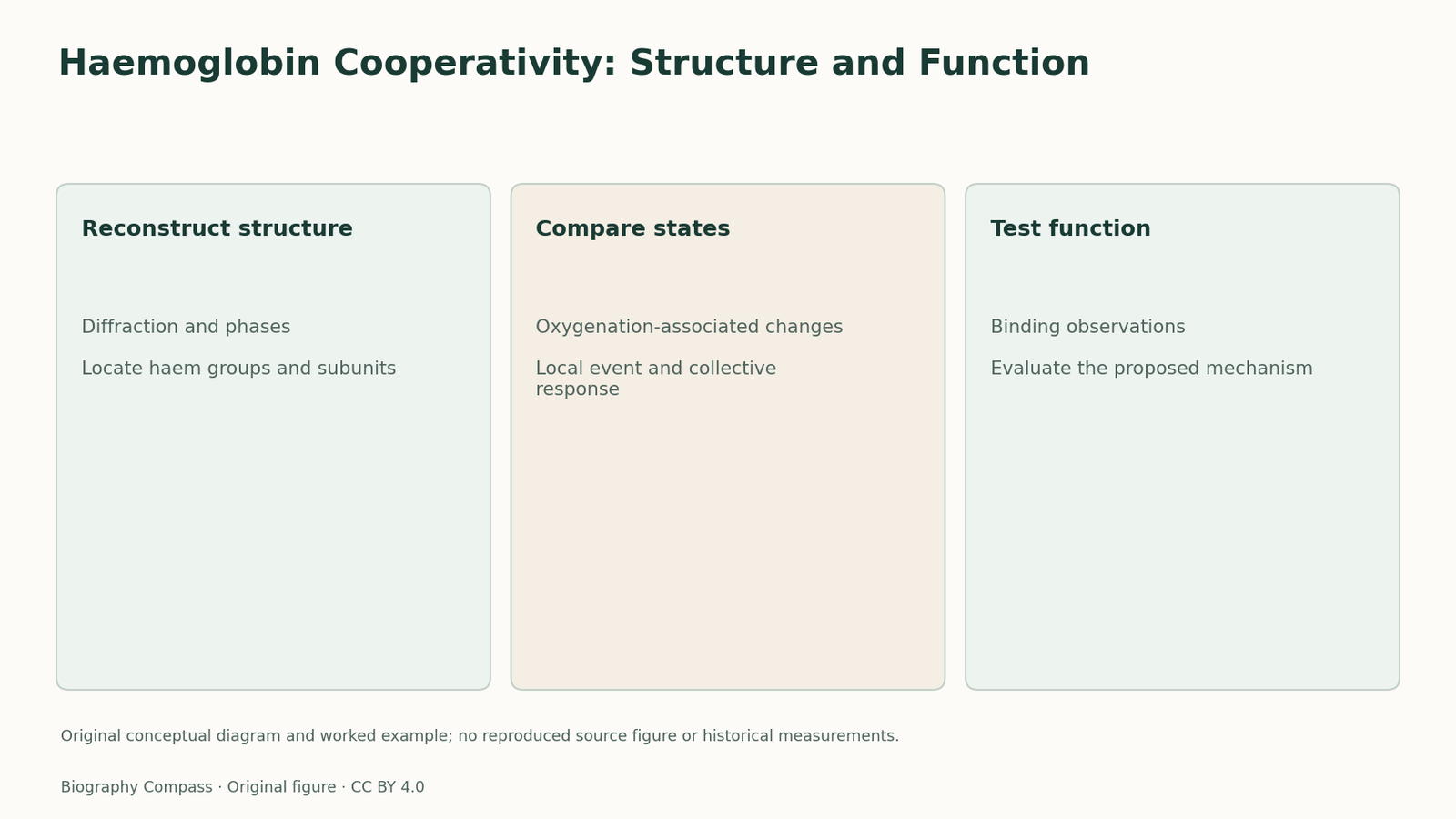
The short answer
How did structural models help explain cooperative oxygen binding without replacing the binding experiments?
Read the evidence
Different records within the same investigation
The following comparison distinguishes the specific questions answered by the inspected records.
| Record or test | What it establishes | What remains different |
|---|---|---|
| Crystallographic structure | Locates molecular features in an experimentally reconstructed arrangement. | Does not alone measure a complete oxygen-binding response. |
| Binding behaviour | Establishes the functional pattern requiring explanation. | Does not by itself locate the structural communication route. |
| Stereochemical mechanism | Connects specific structural changes with a functional interpretation. | Needs comparison with both kinds of evidence. |
A structure addresses an arrangement
Perutz’s 1962 lecture explains how protein crystallography developed from diffraction patterns into molecular models. Heavy-atom derivatives helped address the phase information needed for that reconstruction. The immediate result was an account of where structural features lay in a crystal. This provided an essential starting point for studying haemoglobin, but a structural map and a measurement of oxygen binding were different records. Their relationship became the subject of a further mechanistic programme.
Sources for this section: [2]
The 1970 proposal connected local and collective changes
The 1970 paper described oxygenation-associated shifts involving iron atoms, the surrounding haem groups and the protein subunits. It related these changes to constraints stabilising the oxygen-free form. That was an attempt to explain how binding at one part of a multi-subunit protein could alter the conditions experienced elsewhere. The distinction between a local binding event and a collective structural response is central to the question. It is more specific than saying that a protein changes shape when it works.
Sources for this section: [1]
Compare an architecture with a functional explanation
The earlier crystallographic programme supplied an experimentally grounded architecture; the later paper interpreted how changes within that architecture could contribute to cooperative behaviour. Reading them together makes the chain of reasoning visible. A model needs structural features that can plausibly communicate a change, while binding experiments establish the functional behaviour being explained. Neither record should be substituted for the other. A static view of one state cannot by itself supply an entire binding curve, and a curve cannot directly locate the relevant atomic rearrangements.
An original two-record exercise
Imagine a fictional oxygen-binding protein with two measured structural states and a separate set of binding observations. A student says the second state proves that binding one oxygen molecule always forces every other site to bind immediately. What is missing from that argument? The structural comparison can suggest how sites influence one another, but the size and form of the cooperative effect require functional evidence. Now suppose a mutation preserves the overall fold while changing the binding behaviour. That would motivate a closer test of the proposed communication pathway rather than making the two records incompatible. This exercise contains no historical affinity values or copied protein coordinates. Its supported answer is to connect structural states, a mechanism and functional measurements while identifying the contribution made by each.
Continue the chronology
Related history guides
Myoglobin: From a 6 Å Map to Atomic Detail
Compare Kendrew’s 1958 low-resolution map with the 1960 myoglobin model, using a worked exercise on what improved resolution can reveal about protein structure.
Read the timeline1951The Alpha Helix: A Model Before a Protein Structure
Explore Pauling, Corey and Branson’s 1951 helix proposal, with an original exercise on how molecular geometry constrains a model before a full protein map.
Read the timelineBiography context
Explore United Kingdom through connected lives
Connected biographies
Researchers and related approaches
1914–2002
Max Perutz
Structural biologist and protein crystallographerMax Perutz built a Cambridge programme in protein crystallography, helping reveal haemoglobin’s structure and connect molecular changes with oxygen binding.Read biography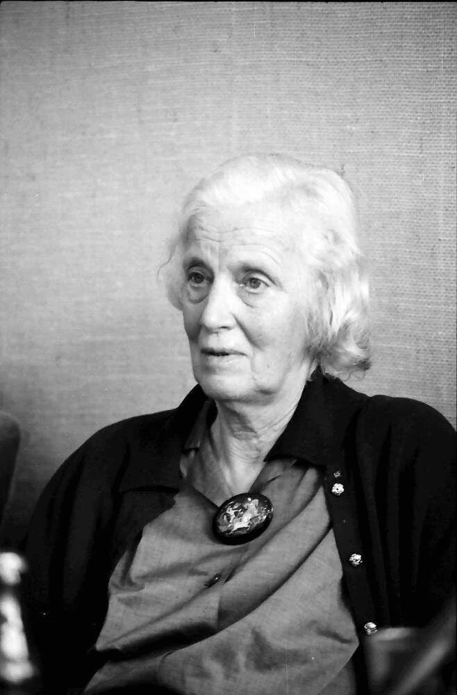
1910–1994
Dorothy Hodgkin
Chemist, crystallographer and university teacherDorothy Hodgkin used X-ray crystallography to investigate the three-dimensional structures of biologically important molecules. Her career joined experimental technique, mathematical interpretation, teaching and international research collaboration.Read biographyQuick answers
Common questions
- Is a structural state an oxygen-binding curve?
- No. They are different records that can be connected through a mechanistic explanation.
- What did the 1970 work add?
- It related oxygenation-associated structural changes to constraints and interactions within haemoglobin.
Primary and institutional sources
How this history was checked
The references below distinguish the original study from later explanations. The worked exercise and comparison are newly written for this article. This article was reviewed on 8 November 2026.