
The short answer
What changed between the 1958 low-resolution myoglobin map and the 1960 atomic model?
Read the evidence
Different records within the same investigation
The following comparison distinguishes the specific questions answered by the inspected records.
| Record or test | What it establishes | What remains different |
|---|---|---|
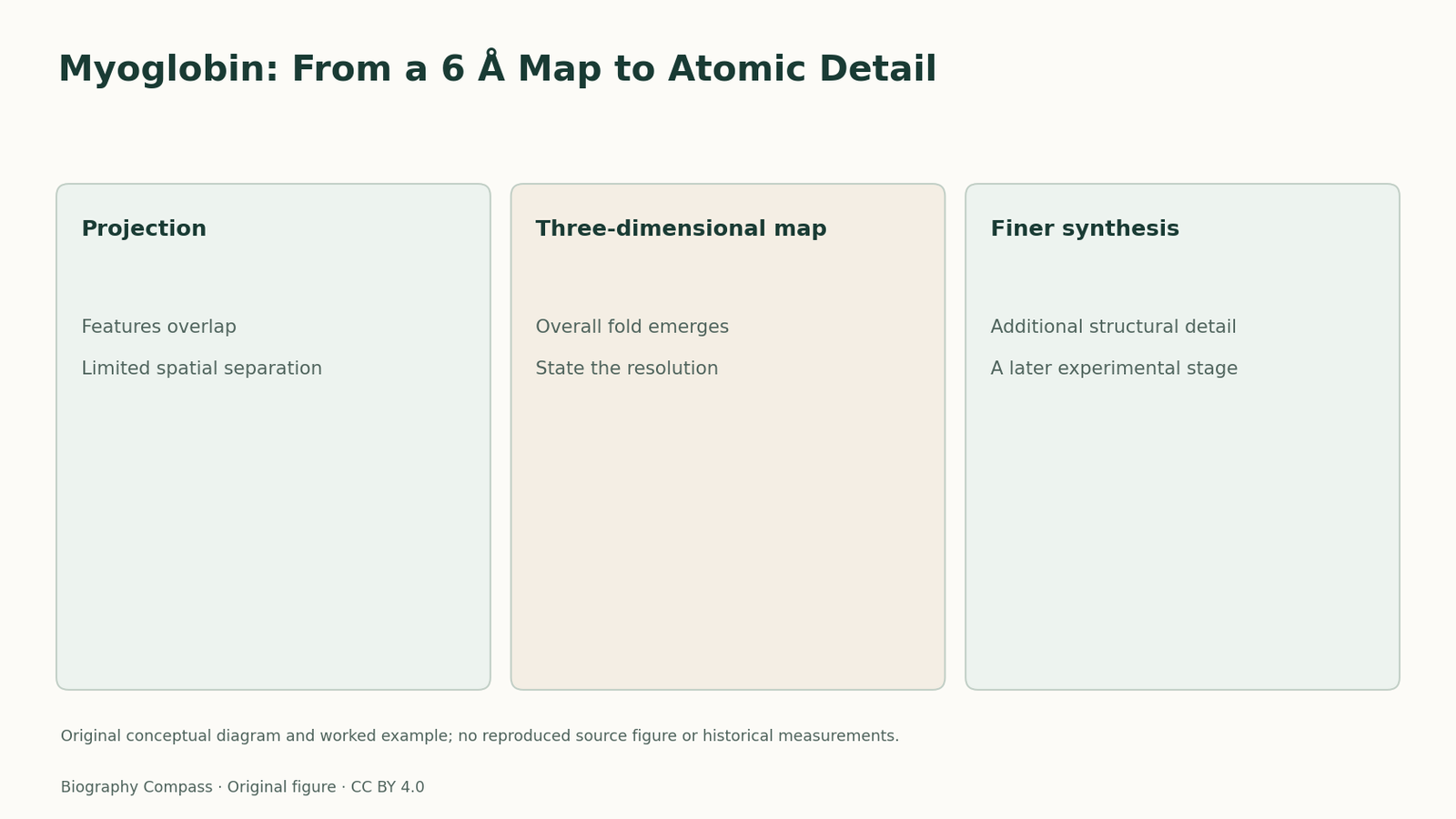
| Two-dimensional projection | Combines features along a viewing direction. | Can obscure the arrangement of a complex protein. |
| Low-resolution three-dimensional map | Reveals broad spatial organisation. | Does not support every fine atomic assignment. |
| Higher-resolution synthesis | Supplies additional structural detail. | Should be credited as a further experimental stage. |
The first goal was a three-dimensional map
Kendrew’s lecture explains why an early two-dimensional projection supplied little useful structural information: features from a complex molecule overlapped in the projection. The programme therefore moved toward a spatial electron-density map. That shift required many more diffraction observations and comparisons among heavy-atom derivatives. It changed the kind of record available to the investigator, from a superimposed view toward a representation in which the protein’s folded arrangement could be followed through space.
Sources for this section: [3]
A 6 Å map and a 2 Å synthesis answer different questions
The 1958 model established the broad folded architecture of myoglobin. The 1960 publication explicitly described a synthesis at2 Å resolution. Improved resolution made finer structural interpretation possible, including a more detailed account of the polypeptide chain and its surroundings. These stages should not be collapsed into a single date followed by the word solved. A molecular structure can be an important advance before every feature is resolved, provided the description matches the actual information available.
Resolution is about information, not presentation
A smoother drawing or a more elaborate physical model does not add experimental detail to a low-resolution map. The improvement depends on the underlying diffraction information and its analysis. Kendrew’s account also shows why material preparation mattered: useful heavy-atom derivatives had to occupy interpretable sites without destroying the comparison. The visual result therefore condensed a large practical research programme. Its explanatory power came from a defined relationship with the measurements, rather than from the apparent realism of the final image. The same protein can therefore have several historically important structural descriptions.
An original map-reading exercise
Imagine two fictional maps of the same folded chain. The first supports the position of several broad segments; the second separates finer features within those segments. A student labels a particular side-chain contact on the first map because it appears in the second. Is that a justified description of the first experiment? No: later knowledge can help interpret an earlier result, but it does not increase the information originally measured. Now ask whether the earlier map was scientifically useless. That conclusion also fails, because an overall fold can answer an important question before atomic detail is available. This exercise uses no myoglobin coordinates or reconstructed diffraction values. Its supported answer distinguishes a major structural discovery from the later refinement of that discovery.
Continue the chronology
Related history guides
Haemoglobin Cooperativity: Structure and Function
Connect Perutz’s structural work with his 1970 account of oxygen binding, using an original exercise to distinguish molecular states from binding measurements.
Read the timeline1951The Alpha Helix: A Model Before a Protein Structure
Explore Pauling, Corey and Branson’s 1951 helix proposal, with an original exercise on how molecular geometry constrains a model before a full protein map.
Read the timelineBiography context
Explore United Kingdom through connected lives
Connected biographies
Researchers and related approaches
1917–1997
John Kendrew
Protein crystallographer and molecular biologistJohn Kendrew moved from wartime operational research to protein crystallography, working with Max Perutz to make myoglobin’s architecture experimentally visible.Read biography1945–
Richard Henderson
Structural biologist and electron-microscopy researcherRichard Henderson moved from physics into structural biology, working with Nigel Unwin and later collaborators to make electron microscopy reveal membrane-protein structure.Read biographyQuick answers
Common questions
- Does a more realistic drawing improve resolution?
- No. Resolution depends on the measured information and analysis, not the visual style of the model.
- Can a low-resolution map still be a major discovery?
- Yes. Establishing the overall fold is a different and valuable achievement from resolving finer atomic detail.
Primary and institutional sources
How this history was checked
The references below distinguish the original study from later explanations. The worked exercise and comparison are newly written for this article. This article was reviewed on 8 November 2026.
- Kendrew and colleagues / Nature, 1958A Three-Dimensional Model of the Myoglobin Molecule Obtained by X-Ray Analysis
- Kendrew and colleagues / Nature, 1960Structure of Myoglobin: A Three-Dimensional Fourier Synthesis at2 Å Resolution
- Nobel Prize OutreachJohn Kendrew — Nobel lecture
- Nobel Prize OutreachJohn Kendrew — Facts
- Nobel Prize OutreachJohn Kendrew — Biographical