
The short answer
Why does changing the angle of an X-ray detector change the wavelength it can register?
Read the evidence
A three-angle prediction
The electron Compton wavelength gives a convenient unit of shift.
| Angle | Cosine term | Shift in units |
|---|---|---|
| 0 degrees | One minus one | Zero |
| 90 degrees | One minus zero | One |
| 180 degrees | One minus negative one | Two |
Where the extra wavelength comes from
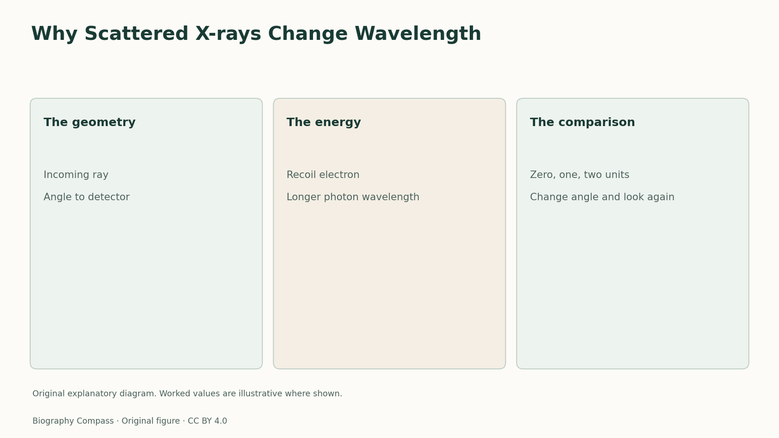
Compton’s 1923 model treated the incoming X-ray quantum as carrying momentum. When it scatters from an electron initially taken to be at rest, the electron can recoil. The scattered radiation then carries less energy than the incident radiation. For a photon, lower energy corresponds to longer wavelength. This makes the altered spectrum a measure of the interaction, because the amount of recoil depends on the geometry. The free-electron picture is an idealisation useful for predicting a shifted component. Conditions in a real target can add intensity at other wavelengths and broaden features.
Read three angles
The shift in the simple model is the electron Compton wavelength multiplied by one minus the cosine of the scattering angle. A detector aligned forward, at zero degrees, corresponds to zero shift. At ninety degrees the change is one electron Compton wavelength. At one hundred and eighty degrees it is twice that scale. Start with a hypothetical incoming wavelength of ten units, with one unit equal to the electron Compton wavelength. The three ideal outgoing wavelengths would be ten, eleven and twelve units. This simple progression gives the angular prediction a form that can be checked independently of the original plot.
Sources for this section: [3]
Look for shifted and unshifted components
A scattering spectrum can show intensity near the incident wavelength as well as a displaced feature. Compton’s lecture discusses evidence for both components. What the simple recoil law predicts is the position of the displaced contribution under its assumptions. Electrons interacting as part of a bound system can contribute to an approximately unshifted signal under suitable conditions. When examining a historical scan, mark the incident wavelength and the angle before identifying peaks. A single spectrum is informative, but the movement of a peak across several angles is more specific evidence for a recoil law.
Separate recoil from fluorescence
Changing the detector angle tests recoil. Changing the element can expose characteristic X-ray radiation associated with that element. Charles Barkla’s work on secondary X-rays helped make the distinction between scattered radiation and element-specific fluorescence a separate experimental question. In Compton’s study the position of the shifted component is tied to the incident wavelength and the scattering angle. In characteristic emission the relevant line energies depend on the emitting material. A spectrum may contain more than one contribution. Attribution therefore depends on how a particular feature responds when one controlled condition changes.
Why a second method helped
A spectrum can compare the position and strength of peaks without showing both products of one interaction in the same record. Wilson’s cloud chamber made recoil-electron paths observable, and coincidence experiments probed event associations. The methods use separate pieces of evidence to test the same physical picture. To compare them, write down what each instrument actually registers. A wavelength analyser measures radiation. A track image reveals the path of a charged particle. Event coincidence can connect observations within the resolution of the equipment. Together they make the explanation stronger without requiring every instrument to measure the same thing.
Continue the chronology
Related history guides
Finding a Neutral Meson through Two Photons
Read the 1950 Berkeley neutral-meson evidence and use a two-photon comparison to understand why coincidences are more informative than isolated counts.
Read the timeline1932–1933How a Lead Plate Identified the Positron
Reconstruct the direction, charge and range checks behind Carl Anderson’s positron identification and see what the lead plate added to a cloud-chamber photograph.
Read the timeline1900s–1910sHow Barkla Separated Characteristic X-rays
Use Barkla’s X-ray evidence to distinguish fluorescence, scattered radiation and recoil shifts, with a three-step comparison of changing conditions.
Read the timelineBiography context
Explore St. Louis and Chicago, United States through connected lives
Connected biographies
Researchers and collaborators
1892–1962
Arthur Compton
Experimental physicist of X-ray scatteringTrace Arthur Compton’s path from Wooster and Princeton to his X-ray research, the 1923 scattering paper and later work on photon and electron evidence.Read biography1877–1944
Charles Glover Barkla
Experimental physicist of X-ray fluorescenceFollow Charles Glover Barkla from Liverpool to Edinburgh and see how his studies of polarization and fluorescence made secondary X-rays an experimental tool.Read biography1862–1942
William Henry Bragg
Physicist and laboratory directorPhysicist and shared Nobel Prize recipient.Read biographyQuick answers
Common questions
- What happens to the X-ray energy?
- The photon loses energy when it transfers some to a recoiling electron, corresponding to a longer wavelength.
- Can a spectrum show an unshifted peak too?
- Yes. The recoil formula describes the shifted component under a free-electron approximation.
Primary and institutional sources
How this history was checked
Dates, legal milestones and historical claims are checked against the official and institutional records below. This article was reviewed on 13 November 2026.