
The short answer
How did the 1975 low-resolution three-dimensional map differ from the 1990 atomic model of a membrane protein?
Read the evidence
Architecture and chemistry require different detail
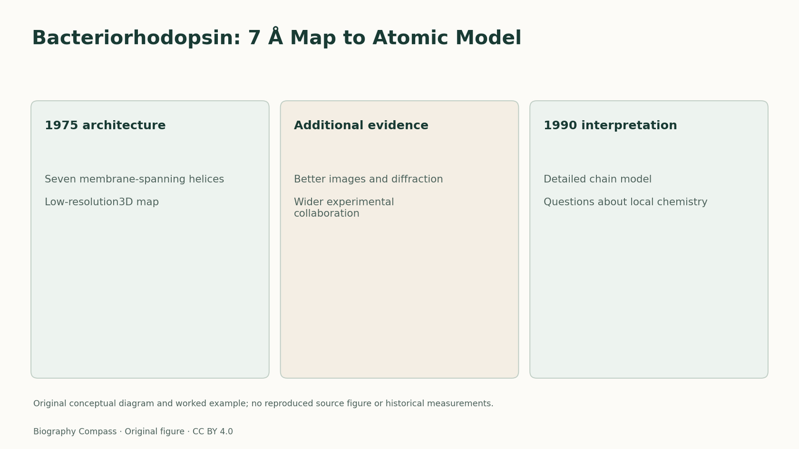
The 1975 and 1990 papers concern the same protein but establish different levels of structural information.
| Record or test | What it establishes | What remains different |
|---|---|---|
| 1975 low-resolution3D model | Reveals a membrane-spanning seven-helix architecture. | Does not locate every side chain. |
| 1990 higher-resolution model | Supports a much more detailed molecular interpretation. | Is not obtained by simply enlarging the earlier drawing. |
| Collaborative development | Improves preparation, imaging and analysis. | A retrospective success does not erase intermediate uncertainty or different contributions. |
The 1975 result already had three dimensions
Henderson and Unwin’s 1975 paper reported a roughly7Å-resolution model of purple membrane obtained by electron microscopy. It identified seven closely packed helical segments running across the membrane. Calling that achievement only a flat image would understate the result. Calling it a fully detailed atomic structure would overstate it. The useful description is a low-resolution three-dimensional architecture, derived from tilted specimens and structural analysis, which showed how the protein traversed the membrane without yet resolving every chemical feature.
Higher resolution changed the questions
The 1990 paper reported a bacteriorhodopsin model based on high-resolution electron cryo-microscopy. The study combined image and diffraction information to support more detailed interpretation of the molecule. At this level, the placement of the polypeptide and side chains became accessible in ways that the earlier helical rods did not permit. Resolution is therefore not only a number attached to a prettier picture. It determines whether a model can answer a broad architectural question or a more specific question about amino-acid arrangement and chemical interactions.
Sources for this section: [2]
The intervening work was experimental, not cosmetic
Henderson’s autobiography describes years of unsuccessful approaches before the group committed to better high-resolution cryo-electron images. Collaborations with other laboratories were part of that development. The later paper’s wider author list reflects those contributions. MRC’s retrospective account places the 1975 result in this longer history, while the original papers retain their different scopes. This prevents a misleading retrospective story in which the atomic model was already present, merely hidden in a low-resolution illustration awaiting someone to sharpen it.
An original resolution-matching exercise
Imagine two invented models. Model A contains seven rods crossing a membrane. Model B also assigns a detailed chain and side-chain positions supported by additional evidence. Ask which can directly address whether the protein crosses the membrane several times, and which is better suited to a question about a particular residue’s local environment. The first question may be addressed by A; the second needs the detail represented by B and appropriate validation. Enlarging A on a screen does not turn it into B. These are conceptual models, not altered source artwork or a reconstruction of the historical maps. The supported answer is to match the scale of the question to the information actually measured, while recognising that both models can be important scientific achievements.
Continue the chronology
Related history guides
Single Particles: Alignment and Classification
Compare image alignment with particle classification in Frank and van Heel’s early work, using an original exercise that separates orientation from molecular state.
Read the timeline1989; later review 2015A Single Molecule: Signal and Background
Examine Moerner and Kador’s 1989 absorption experiment, distinguishing spectral selection, background rejection and later fluorescence-based imaging.
Read the timelineBiography context
Explore United Kingdom through connected lives
Connected biographies
Researchers and related approaches
1945–
Richard Henderson
Structural biologist and electron-microscopy researcherRichard Henderson moved from physics into structural biology, working with Nigel Unwin and later collaborators to make electron microscopy reveal membrane-protein structure.Read biography1926–2018
Aaron Klug
Structural biologist and physical chemistAaron Klug used physics, crystallography and electron microscopy to investigate biological structures, later directing Cambridge’s MRC Laboratory of Molecular Biology.Read biographyQuick answers
Common questions
- Why is it wrong to call the 1975 model only a two-dimensional projection?
- The Nature report explicitly presented a three-dimensional low-resolution model from electron-microscopy work.
- Does enlarging a map improve its scientific resolution?
- No. Additional display size cannot create structural information that was not measured.
Primary and institutional sources
How this history was checked
The original 1975 and 1990 records are compared with Henderson’s account and MRC’s institutional retrospective. No source structural images are reproduced. This article was reviewed on 7 November 2026.
- Richard Henderson and Nigel Unwin / NatureThree-dimensional model of purple membrane obtained by electron microscopy
- Henderson, Baldwin, Ceska, Zemlin, Beckmann and Downing / Journal of Molecular BiologyModel for the structure of bacteriorhodopsin based on high-resolution electron cryo-microscopy
- MRC Laboratory of Molecular BiologyThe structure of bacteriorhodopsin50years on
- Nobel Prize OutreachRichard Henderson — Facts
- Nobel Prize OutreachRichard Henderson — Biographical