
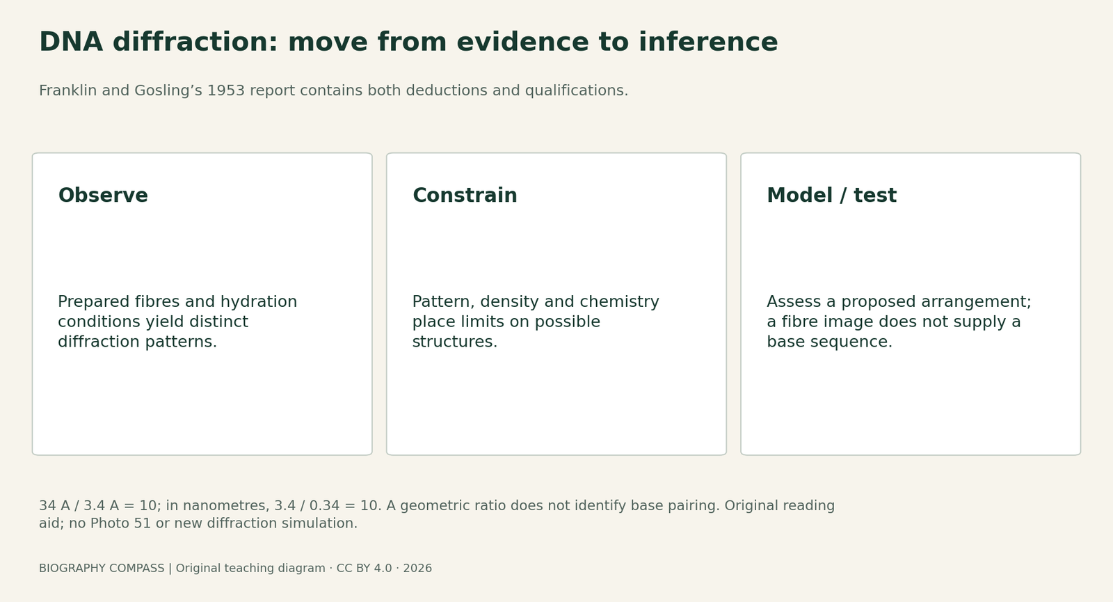
The short answer
Photo 51 is famous, but a photograph, a structural inference and a complete molecular model are different things. Read the April 1953 records together to see what the experiments supported, what remained conditional and how several teams contributed.
Original source comparison
Read the record by its job
Match the claim to the record that can support it.
| Country | What it supplies | What it does not establish alone |
|---|---|---|
| Franklin and Gosling | Hydration, diffraction and qualified structural deductions | A full base sequence or complete atomic model |
| Wilkins, Stokes and Wilson | Experimental constraints and a separate team account | Sole authorship of all King’s observations |
| Watson and Crick | A two-chain model and base-pairing proposal | Independent performance of all cited experiments |
| Later institutional history | Context about appointments and access to results | A substitute for the exact wording of the 1953 papers |
Chronology
A timeline of the turning points
Franklin at King’s
Joined the MRC Biophysics Unit and worked with Gosling.
B-form image
Photo 51 records prepared B-form material.
MRC visit
A report became a separate route for experimental information.
Three papers
The model and experimental discussions appeared together.
Begin with the experiment, not the familiar model
Franklin and Gosling studied prepared sodium DNA fibres under controlled conditions. Their April 1953 paper distinguishes A and B diffraction patterns and a reversible change associated with hydration. Photo 51 records B-form material. It is not a direct picture of two visible spiral rails: interpreting the pattern requires a relationship between scattering, the specimen and a possible structure.
The original article explicitly stops short of treating the X-ray evidence alone as direct proof of a helix. Its reasoning combines the pattern with other observations and structural arguments. Reading that qualification changes the question from “Who recognised the picture?” to “Which measurements constrain which part of a proposed model?”
Three papers make different contributions
Nature’s issue of 25 April 1953 placed Watson and Crick’s model on pages 737–738, Wilkins, Stokes and Wilson’s experimental paper on pages 738–740, and Franklin and Gosling’s report on pages 740–741. The author lists are part of the evidence: Raymond Gosling, Alec Stokes and Herbert Wilson should remain visible alongside the more familiar names.
A useful reading method is to mark a statement as an observation, an inference or a proposed model. Watson and Crick’s paper sets out a double-chain arrangement and base-pairing argument. The experimental papers address diffraction observations and structural constraints. Placing them together makes it possible to compare claims without pretending that each paper contains the same experiment or that publication order measures each person’s contribution.
Sources for this section: [1]
What the fibre data can and cannot decide
Franklin and Gosling report a repeat of 34 Å along the fibre axis and a strong reflection at 3.4 Å. Their discussion combines such quantities with density, symmetry and chemical considerations. It argues for phosphates on the outside and a probable two-chain structural unit while retaining qualifications. A model that ignored water content or placed the phosphates in a dense central core would have to answer those constraints.
The letter does not supply the nucleotide sequence of the specimen. Nor does an X-shaped pattern by itself establish base-pair identities, every atomic coordinate or a biological replication mechanism. Those are different questions. Separating them is not a judgement that the photograph was unimportant; it is how its actual evidential value becomes precise.
Sources for this section: [1]
Access and attribution are separate from structural validity
King’s describes two routes by which unpublished experimental information reached Cambridge: Wilkins showed Watson a B-form image, and an account of the MRC’s December 1952 visit was subsequently available there. These should not be collapsed into one event. The institutional history also records earlier work by Wilkins and Gosling and a role for Stokes in interpreting helical diffraction.
The printed papers let a reader check author lists, acknowledgements and claims. They cannot reconstruct every private conversation or determine an alternative Nobel outcome. A fair source comparison therefore distinguishes what a surviving document states, how a later account interprets access, and what remains unknown. The later death-date error in one King’s overview is another reason to cross-check individual statements rather than treating an institutional page as infallible.
An original source-reading exercise
Work with these four claims: A, the specimen showed different diffraction patterns at different hydration conditions; B, the fibre image itself displays a readable DNA sequence; C, a repeat of 34 Å divided by a 3.4 Å spacing gives a ratio of 10; D, the number 10 alone proves which bases pair. Classify each as supported observation, valid limited calculation or unsupported inference. Use the paper’s qualifications as well as its positive conclusions.
Answers: A is a supported experimental observation. B is unsupported: diffraction is not a sequence readout. C is a valid numerical ratio, 34/3.4 = 10, for the stated quantities; converting both to nanometres gives 3.4/0.34 and preserves that ratio. D is unsupported because a geometric ratio does not identify the chemical pairing scheme. The exercise is newly written and uses no newly measured diffraction data.
Extension: write a caption naming Franklin and Gosling, identifying B-form fibre diffraction and stating that interpretation used additional evidence. Explain why a claim that the image alone proves every feature of DNA would be stronger than the cited experimental report. A good answer names a specific missing inference rather than substituting a different heroic story.
Continue the chronology
Related history guides
Connected biographies
People and research
1920–1958
Rosalind Franklin
Physical chemist and X-ray crystallographerRosalind Franklin investigated the structure of matter through careful preparation, diffraction measurements and mathematical analysis. Her DNA research with Raymond Gosling was one part of a career that also made substantial contributions to carbon and virus studies.Read biography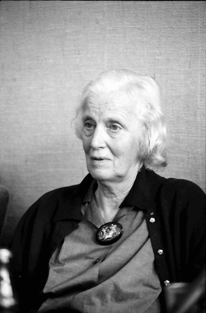
1910–1994
Dorothy Hodgkin
Chemist, crystallographer and university teacherDorothy Hodgkin used X-ray crystallography to investigate the three-dimensional structures of biologically important molecules. Her career joined experimental technique, mathematical interpretation, teaching and international research collaboration.Read biography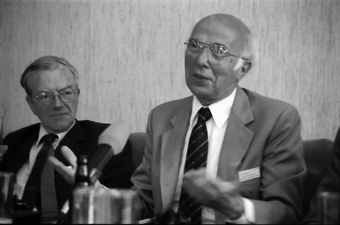
1916–2004
Maurice Wilkins
Physicist and molecular biophysicistMaurice Wilkins moved from solid-state physics and wartime research into molecular biophysics at King’s College London. His DNA work combined instruments, diffraction experiments and years of structural investigation, within a discovery involving several laboratories and collaborators.Read biographyQuick answers
Common questions
- Was Photo 51 a direct photograph of two DNA strands?
- No. It is a diffraction pattern from prepared B-form DNA fibres. Structural information is inferred from the pattern together with other evidence.
- Who authored the three April 1953 papers?
- Watson and Crick wrote the model paper; Wilkins, Stokes and Wilson wrote one experimental paper; Franklin and Gosling wrote the other.
- Are the exercise and diagram original experimental results?
- No. They are newly written aids for reading the historical sources. The numerical ratio uses quantities stated in the original paper.
Primary and institutional sources
How this history was checked
Original research records support the historical discussion. The comparisons, questions and figures are newly written learning aids; they are not new experimental results or reproductions of archival images. This article was reviewed on 27 October 2026.